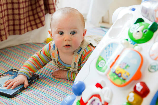
Welcome!We are a group of researchers interested in the development of infants in their first year of life. We study what infants are thinking and what they are learning.
By studying healthy infants, we aim to improve understanding of brain development during the first year and to improve diagnoses and therapies for infants who have a difficult start in life. |
|
Deep Learning as a Model of Infant Development
|
By the time infants reach a few years old, they have developed many complex cognitive functions; for example they can walk, talk and solve problems. These cognitive functions do not suddenly appear, but instead they emerge from a combination of innate mechanisms and life experience. Our group is trying to understand how these cognitive functions develop over the course of an infant's first year of life.
Of course, it is hard to imagine what an infant's mind is actually like, and the power of neuroscience methods such as MRI can only tell us so much. Therefore, our approach is to harness the benefit of using computational models for development. Deep learning, the technology underlying modern artificial intelligence, has recently made strides in modelling the functions of the adult brain. In our "Foundations of Cognition" project, we aim to test whether the mechanisms of learning in artificial neural networks can provide a testable model of how human infants learn. The "Foundations of Cognition" is supported by the European Research Council, and runs from January 2019 - December 2023. To participate, hear more about the goals and discoveries of the project, or opportunities to help shape it, please join our mailing list. See our Vacancies page for opportunities, or if you are interested in joining our team and have experience in deep learning with PyTorch or TensorFlow, please do get in touch. |

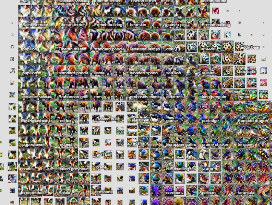
Visualizing how artificial neural networks represent objects (distil.pub, the activation atlas)
|
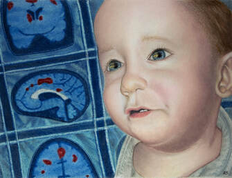
Infant Brain and MindWe study what infants are thinking and what they are learning. This research will help us know ourselves and our children, and help us understand how the development of brain functions goes awry following perinatal brain injury.
|
|
|
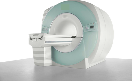
Neuroimaging MethodsNeuroimaging with magnetic resonance imaging is a powerful tool to help us understand the brain.
We develop methods to improve brain scanning, by acquiring better data, and improve what we can learn about the brain with new analysis and modelling methods. |